
Monitoring of electrocardiogram (ECG) for bipolar limb leads l1,l2 and l3.
Introduction
An ECG is a series of waves and deflections recording the heart’s electrical activity from a certain view. Each part of wave corresponds to particular action in heart as shown. Many views, each called a lead, monitor voltage changes between electrodes placed in different position on the body.
The interior of the cell membrane is considered to be negative with respect to outside during resting conditions. When an electric impulse is generated in the heart, the interior part becomes positive with respect to the exterior. This change of polarity is called depolarization. After depolarization the cell comes back to its original state. This phenomenon is called repolarization. The ECG records the electrical signal of the heart as the muscle depolarize (contract) and repolarize.
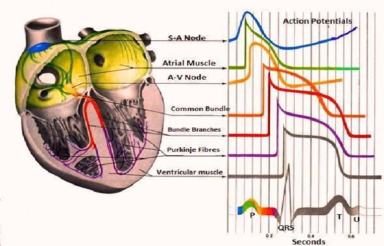
Figure-1: Leads Position
The SA node is the basic, natural cardiac pacemaker that triggers its own train of action potentials. The action potential of the SA node propagates through the rest of the heart, causing a particular pattern of excitation and contraction. The sequence of events and waves in cardiac cycle is as follows:
- • The SA node fires.
• Electrical activity is propagated through the atrial musculature at comparatively low rates, causing slow –moving depolarization (contraction) of the atria. This results in the P wave in the ECG. Due to the slow contraction of the atria and their small size, the P wave is a slow, low amplitude wave, with an amplitude of about 0.1-0.2 mV and a duration of about 60-80 ms.
• The excitation wave faces a propagation delay at the atrio- ventricular (AV) node, which results in a normally iso-electric segment of about 60-80 ms after the P wave in ECG, known as the PQ segment. The pause assists in the completion of the transfer of blood from the atria to the ventricles.
• The bundle, the bundle branch and the purkinje system of specialized conduction fibers propagate the stimulus to the ventricles at the high rate.
• The wave of stimulus spread from the apex of the heart upwards, causing rapid depolarization (contraction) of the ventricles. This results in the QRS wave of the ECG- a sharp triphasic wave of about 1mV amplitude and 80 ms duration.
• Ventricles muscle cells possess a relatively long action potential duration of about 300-350 ms. The plateau of the action potential causes normally iso-electric segment of about of the 100- 120 ms after the QRS, known as the ST segment.
• Repolarization (relaxation) of the ventricles causes the slow T wave, with an amplitude of 0.1-0.3 mV and duration of 120-160 ms.
ECG Electrode Placement & ECG Leads –
12 Lead ECG configuration:-
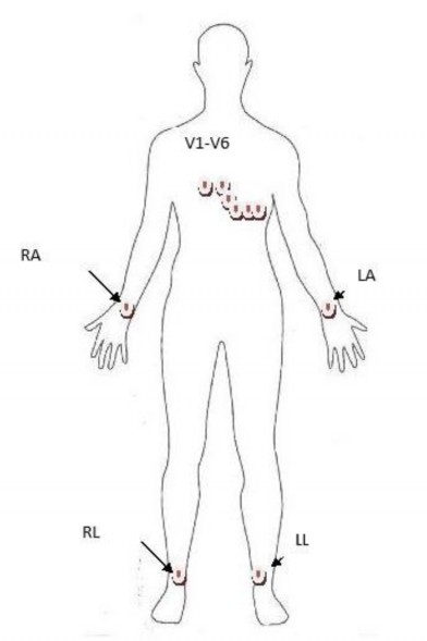
Figure-2: 12 Lead ECG configuration
In ECG lead refers to the voltage between two electrodes. In 12 Lead ECG total 10 surface electrodes are attached to various specified positions on the body as shown in the figure 2. A suitable gel is used to provide impedance match between the electrodes and the skin. The 10 electrodes are classified as: 4 limb electrodes, Right Arm (RA), Left Arm (LA), Left Leg (LL) and Right Leg (RL); and 6 chest electrodes, V1-V6.
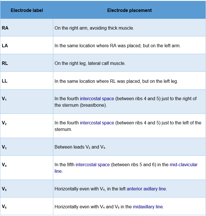
Figure-3: Leads Position
A total of 12 Leads are derived from these 10 electrodes. These 12 leads are classified as 3 limb leads, 3 augmented limb leads and 6 precordial leads. Out of this 12 leads, limb leads are bipolar while all other leads are unipolar leads. The definition for all 12 leads is as follows:-
• Lead I – is the signal between negative RA and positive LA electrodes.
• Lead II – is the signal between negative RA and positive LL electrodes.
• Lead III – is the signal between negative LA and positive LL electrodes.
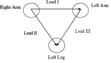
Figure-4: Einthovens Triangle
These three limb leads form the points of what is known as Einthoven's triangle as shown in figure 3, which is the theoretical triangle drawn around the heart with heart at center. According to Einthoven’s law,
Lead I – Lead II + Lead III = 0.
• Lead aVR or Augmented vector right – is the signal between the positive electrode on the right arm and the negative electrode which is a combination of the left arm electrode and the left leg electrode.
• Lead aVL or augmented vector left – is the signal between the positive electrode on the left arm and the negative electrode which is a combination of the right arm electrode and the left leg electrode.
• Lead aVF or Augmented vector foot – is the signal between the positive electrode on the left leg and the negative electrode which is a combination of the right arm electrode and the left arm electrode.
• Precordial leads V1-V6 – is the signal between the corresponding positive V1-V6 electrode on the chest and the negative electrode formed by the Wilson terminal obtained by adding three limb leads.
• (Note: Wilson terminal is formed by the average of the 3 limb leads and approximate ground. This is possible because of Einthoven's Law which states that I + (-II) + III = 0.)